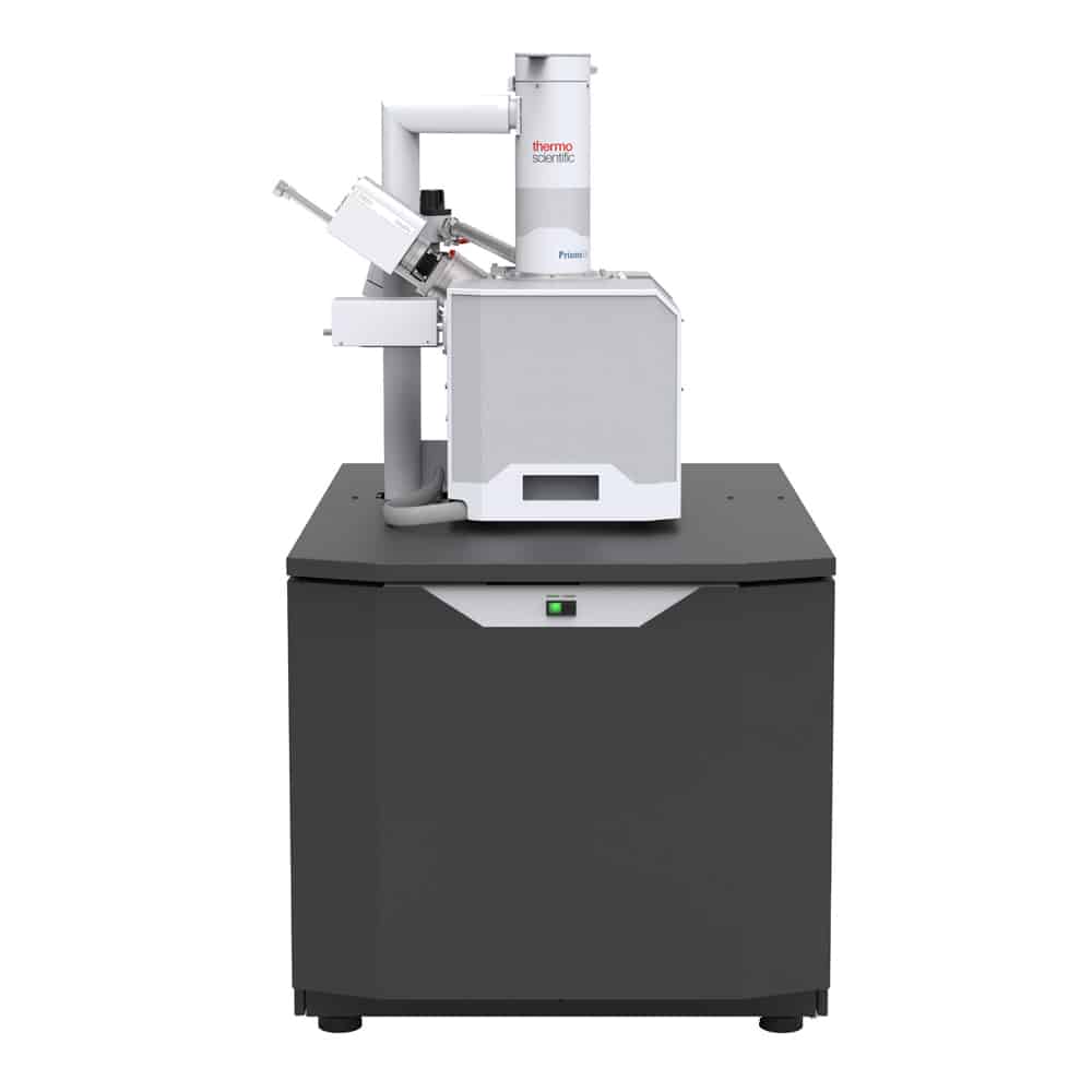
Scanning electron microscope
Sku: HTI04017
Short description: A scanning electron microscope (SEM) is a type of electron microscope that produces images of a sample by scanning the surface with a focused beam of electrons.
Product information
This post is also available in: Tiếng Việt (Vietnamese)
A scanning electron microscope (SEM) is a type of electron microscope that produces images of a sample by scanning the surface with a focused beam of electrons. The electrons interact with atoms in the sample, producing various signals that contain information about the surface topography and composition of the sample. The electron beam is scanned in a raster scan pattern, and the position of the beam is combined with the intensity of the detected signal to produce an image.
Key benefits
- Live composition-based image coloring for the most intuitive elemental analysis with optional ColorSEM Technology and integrated EDS. Speed up your work and obtain the most complete sample information with always-on analysis.
- Minimize sample preparation time: low vacuum and ESEM capability enable charge-free imaging and analysis of nonconductive and/or hydrated specimens..
- Excellent image quality at low kV and low vacuum thanks to flexible vacuum modes including through-the-lens differential pumping. Simultaneous SE and BSE imaging in every mode of operation.
- In-situ study of materials in their natural state: With Prisma E’s ESEM mode, samples can be imaged even if they are hot, dirty, outgassing or wet.
- Excellent analytical capabilities with a chamber that allows 3 simultaneous EDS detectors, EDS ports that are 180° opposite, WDS, coplanar EDS/EBSD and high quality charge free EDS and EBSD in low vacuum.
- Easy to use, intuitive software with User Guidance and Undo functionality makes highly effective operation possible for novice users, while enabling experts to do their work faster and with fewer mouse clicks.


